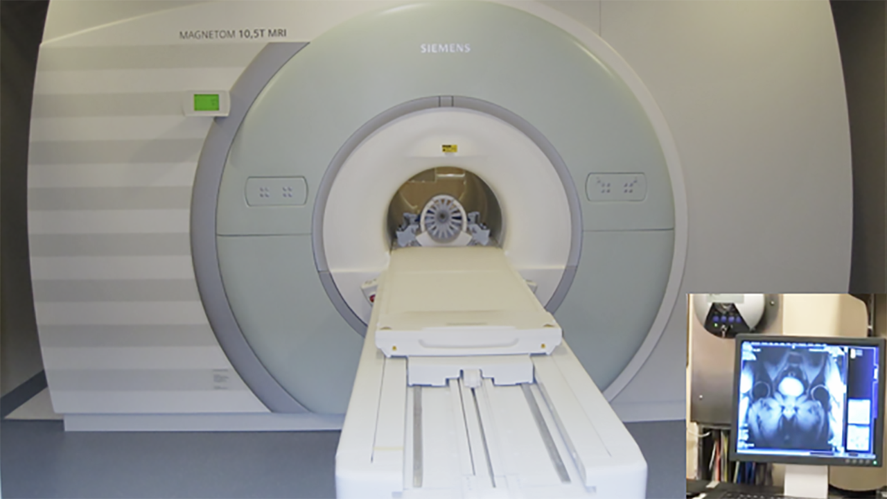
The 10.5-Tesla imaging magnet at the Center for Magnetic Resonance Research. Photo courtesy CMRR.
Scientists at the University of Minnesota have become the first in the world to perform magnetic resonance imaging of the human body at 10.5 Tesla—a magnetic field strength 10 times greater than a standard MRI and topping even the most advanced scanners elsewhere in the world.
In December, researchers in the Center for Magnetic Resonance Research successfully conducted the first scans with the center’s 10.5-Tesla, whole-body imaging magnet. The 110-ton magnet promises to produce scans at a finer level of detail, bringing new capabilities to scientists studying how the body works. In turn, these discoveries may help physicians more precisely choose the best treatment options for a wide range of diseases, such as Alzheimer’s, heart disease, diabetes, and cancer.
CMRR, home to some of the most advanced magnetic resonance instrumentation in the world, is a leader in advanced biomedical imaging. The technologies used at the center are some of the most important tools available for understanding the structure and workings of organs in the body, which is important for both basic and translational research.
Kamil Ugurbil, Ph.D., director of CMRR and professor of medicine, neurosciences and radiology at the U’s Medical School, said the scans were a gratifying milestone after years of preparation.
“It feels great,” Ugurbil said. “We are all excited about it. It’s been a long road leading up to this point.”
The 10.5-Tesla magnet project was first conceived of in 2008, and shortly after CMRR researchers received an $8 million National Institutes of Health (NIH) grant to fund the construction of the magnet. In the years that followed, the magnet was built in England, shipped across the Atlantic Ocean through the Great Lakes to Duluth, and finally brought over land on a specialized trailer to the Center for Magnetic Resonance Research building on the Twin Cities campus.
The CMRR team then spent years preparing the magnet for operation, including cooling the conductors below liquid Helium temperature (2.6 Kelvin or about -455 degrees Fahrenheit/-270 degrees Celsius), configuring its complex electronics, and conducting animal studies to ensure safety for human use. After applying for and earning approval from the US Food and Drug Administration, they are now able to start the first human tests.
Bringing the Brain into Sharper Focus
Going forward, the CMRR team will further develop the capabilities of the 10.5-Tesla magnet and its supporting hardware. While the magnet is capable of imaging the entire body, a major focus is on the brain. Much of what we currently know about brain circuitry comes from the Human Connectome Project, a collaborative national effort that included the work of CMRR and changed the landscape of how scientists understand the brain.
In fall 2017, the CMRR team received a five-year, $9.7 million NIH Brain Initiative grant to pursue the next generation of brain imaging with the magnet. While the main goal of this grant is to conduct basic research into the workings of the brain and to reach currently unavailable spatial scales in human brain studies, going from ensembles of few thousand neurons representing elementary circuits to whole brain function and structural connectivity, the research also holds potential for better understanding and treating neurological diseases, such as Alzheimer’s and Parkinson’s.
Given that most of the Human Connectome Project was conducted with a 3-Tesla magnet, Ugurbil said, the much stronger 10.5-Tesla magnet promises to be the right tool for unlocking new discoveries and keeping the U of M at the forefront of neuroscience research.
“This is an instrument with which we want to push the boundaries of imaging brain function,” he said.
Watch a time lapse video produced by U of M Health Talk of the 10.5-Tesla magnet’s installation at CMRR: