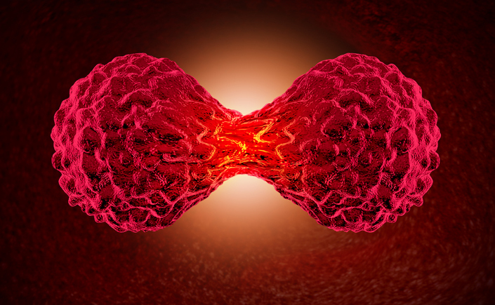
When a cell divides by the process of mitosis, its chromosomes perform a well-choreographed ballet.
First, each makes a copy of itself. The copies, held together as pairs, line up in a ring around the middle of a football-shaped structure called the mitotic spindle. The chromosomes are then pulled apart, with the members of each pair migrating to opposite poles of the spindle. This creates two sets of chromosomes, one in each of the two daughter cells.
But if any chromosomes “lag”—that is, fail to line up and segregate properly—the daughter cells end up with too many or too few chromosomes. This condition, called aneuploidy, is a common feature in cancer development.
Now, researchers at the University of Minnesota’s Hormel Institute have discovered the basis for a “fail-safe” signaling mechanism that monitors chromosome movements and alerts both daughter cells when they are headed for aneuploidy. This begins a process that climaxes with the daughter cells self-destructing.
Published in Nature Cell Biology, the discovery will be invaluable in finding means of healing the aneuploidy detection process when it is defective.
“This study highlights the continued need for research in basic cell biology,” says Edward Hinchcliffe, leader of Hormel’s Cellular Dynamics section, who led the research with Zigang Dong, Hormel Institute executive director.
Threads and Spools
Few structures in nature are as delicate as the threadlike strands of DNA on which life depends. But nature doesn’t let them float unprotected inside the nucleus. Instead, at regular intervals each thread of DNA wraps around structures that may be likened to spools. These spools consist of proteins called histones, which stabilize the DNA and help regulate gene activity. Together, histones and DNA strands form the structures called chromosomes.
The researchers cooled and rewarmed monkey kidney cells, a treatment that induces a high frequency of aneuploidy during cell division. They then studied the differences between cells whose chromosomes “missegregated,” resulting in aneuploidy, and cells that divided normally.
The scientists found that the key lay with one particular histone, known as H3.3. During normal mitosis, the dividing cell “labeled” all chromosomes with phosphate near the region where the two copies were held together. The phosphate label was attached to one particular amino acid in the H3.3 molecule and was removed as the chromosomes were pulled apart.
In cells where aneuploidy developed, phosphate was added in the same manner to the lagging chromosome(s). But not at the connecting region–only along the arms of the chromosomes. This pattern of phosphate labeling quickly spread to all the chromosomes involved in the cell division and persisted in both daughter cells for hours.
Outliers Get Noticed
The unique labeling pattern occurred whenever a chromosome got separated from the others. It was strongest when a single chromosome went astray and weaker when lagging chromosomes clustered together, suggesting that a lack of proximity to other chromosomes was the trigger.
In daughter cells with aneuploidy, the phosphate label lasted long enough for a well-known gene, called p53, to be activated. This gene works to shut down further cell division until the cells can self-destruct–a potent cancer prevention mechanism because aneuploid cells are more prone to turning cancerous. The researchers confirmed their observation by blocking phosphate labeling of chromosomes during mitosis; in that case, aneuploid daughter cells didn’t activate p53.
“This provides a direct biochemical link between chromosome missegregation and triggering of the aneuploidy fail-safe, which depends on the activation of p53,” says Hinchcliffe.
The researchers posit that when histone H3.3 is labeled with phosphate along chromosomal arms, it can set in motion the process that activates p53. They found the same patterns of phosphate labeling in human cells, which also have the p53 gene. They note that in some cancers, including a pediatric brain cancer and certain bone and breast cancers, the H3.3 molecule is mutated in ways that likely prevent phosphate labeling.
The team continues to search for molecular players involved in aneuploidy monitoring and suppression.
On June 1, 2016, the Hormel Institute celebrated a two-year building project that doubled its size and will greatly expand its cancer research capabilities. Among the new labs is one housing a high-resolution electron microscope that captures 2-D and 3-D images using cryo-electron microscopy. Article Published in June 2016.